


Although a thyroid ultrasound is usually not part of a routine physical exam, its diagnostic value is significant.
This small, butterfly-shaped organ located in the front of the neck is highly sensitive to the stressful conditions we are, unfortunately, surrounded by every day. Living in a high-pressure, fast-paced society subjects our bodies to chronic stress, which directly disrupts our delicate hormonal balance.
As a result, thyroid function is often compromised before we even notice the first symptoms.
What to Expect: Do I Need to Prepare?
The good news is that a thyroid ultrasound requires absolutely no prior preparation.
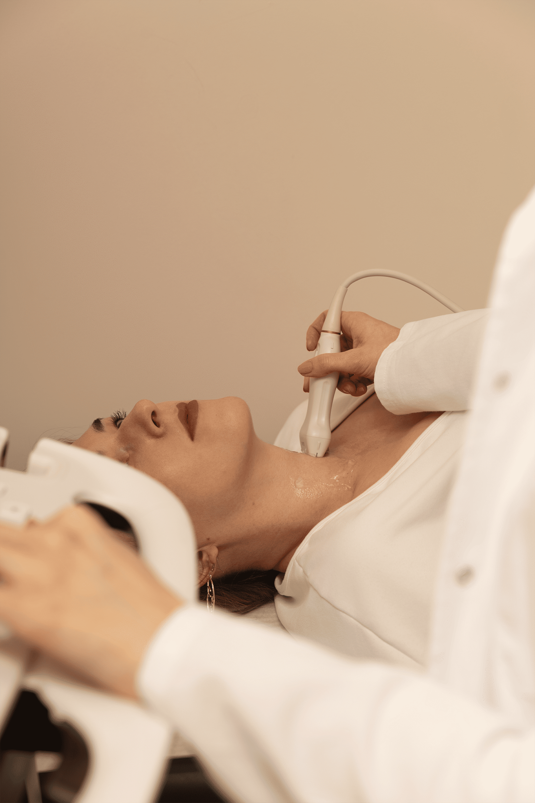
The examination is completely painless, comfortable, and non-invasive. The patient lies comfortably on their back with their head slightly tilted back, while the specialist applies a water-based gel to the neck area and glides the ultrasound probe over it.
The entire procedure typically takes between 15 and 30 minutes, after which you can immediately return to your daily activities.
Cysts and Nodules: Findings That Shouldn't Immediately Scare You
Many patients experience fear when told during an exam that they have “changes” or lesions on their thyroid gland.
“The appearance of thyroid cysts is extremely common and, in the vast majority of cases, is no cause for concern,” explains our endocrinologist, Dr. Neda Crnjak.
The primary difference between these changes lies in their composition:
- Cysts are mostly benign, fluid-filled formations;
- Nodules are solid masses of tissue that can have benign or malignant characteristics;
- Complex (mixed) nodules also exist, containing both fluid and solid components.
The size of the cyst or nodule is not the only determining factor for further diagnostic steps. Sometimes, cysts or nodules can be 2 to 3 centimeters (or larger), and standard medical practice only requires regular ultrasound monitoring every 6 to 12 months.
Conversely, they can be much smaller yet still require further treatment. To accurately assess the severity and nature of the nodules, doctors use internationally recognized TI-RADS (Thyroid Imaging Reporting and Data System) classification. This scoring system, based on specific ultrasound features, allows the physician to make a reliable decision on whether the finding simply needs to be monitored or if a fine-needle aspiration (FNA) biopsy is necessary for a more detailed analysis.
When Even Benign Changes Require Treatment
Even when cysts are entirely benign, their size can begin to cause mechanical obstructions in the neck.
Patients with large cysts or nodules frequently report an uncomfortable “lump in the throat” sensation, difficulty swallowing (dysphagia), or hoarseness due to the pressure the mass exerts on surrounding tissues (the trachea and larynx) and the recurrent laryngeal nerve, which controls the vocal cords.
In such situations, the ultrasound plays a crucial role in helping the doctor determine the best therapeutic approach. Based on ultrasound imaging, a decision is made on how to resolve the issue:
- Fluid evacuation (aspiration): Draining the fluid via a needle;
- Sclerotherapy: Injecting a substance that collapses the cyst walls and prevents it from refilling;
Surgical removal: Recommended when minimally invasive options are not viable.
Structure Without Function Doesn't Tell the Whole Story
The primary purpose of an ultrasound is to provide the doctor with a detailed look at the physical structure of the thyroid gland. It clearly visualizes its homogeneity, the presence and dimensions of any cysts or nodules, as well as vascularization (blood flow to the tissue).
However, the structural image alone cannot tell us how the organ is actually functioning. This is why an ultrasound must always be combined with laboratory analyses of thyroid hormones (such as TSH, FT3, and FT4) and thyroid antibodies. Only when a specialist pairs the insight into the gland’s physical structure with an analysis of its hormonal function can a precise and complete clinical picture be formed. These two components are equally important and inseparable.
Patients requiring regular ultrasound and laboratory diagnostics are most commonly dealing with hypothyroidism (an underactive thyroid gland), which, alongside diabetes and osteoporosis, is one of the three most common endocrinopathies.
Hashimoto’s thyroiditis holds a special place in diagnostics: it is a frequent autoimmune disease that can be clearly seen on an ultrasound as a specific inflammatory change with a disrupted, heterogeneous tissue structure.
Schedule Your Check-Up in Time
The overarching goal of a thyroid ultrasound is the careful monitoring of the characteristics and dimensions of nodules and cysts, preventing them from developing into serious, health-threatening conditions. If you are exposed to high levels of stress on a daily basis, experience chronic fatigue, or suspect a hormonal imbalance, take responsibility for your health.
Schedule an appointment with an endocrinologist at Primea. Our team of experts is at your disposal to thoroughly examine your body’s condition and provide you with peace of mind through timely and precise diagnostics.